2017 Nobel Prize in Chemistry awarded to cryo-electron microscopy developers
What's the story
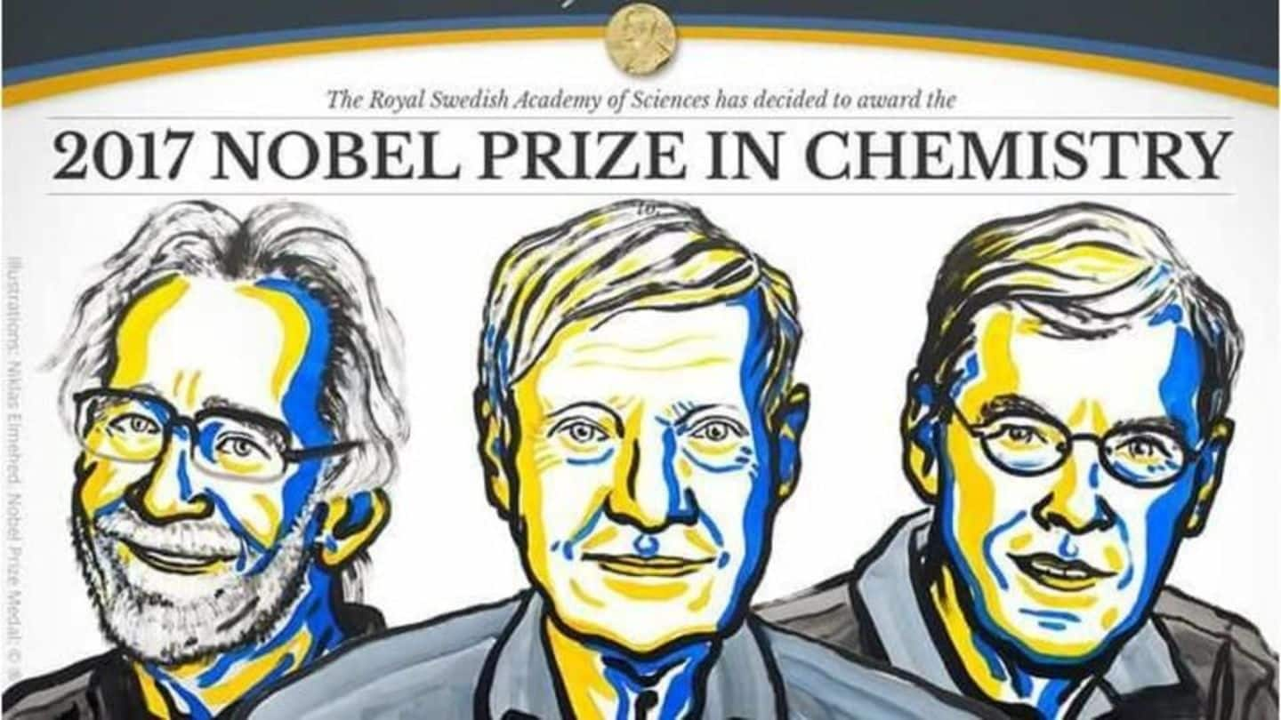
The 2017 Nobel Prize in Chemistry has been awarded to three scientists who developed the cryo-electron microscopy technique to study structures of biomolecules in high-resolution for the first time. The Royal Swedish Academy of Sciences in Stockholm awarded the prize to Jacques Dubochet of Switzerland, Joachim Frank of the US, and Richard Henderson of the UK. Their contribution has transformed the field of biochemistry.
Background
The Nobel Prize in Chemistry
Scientists in various fields of chemistry are awarded the Nobel Prize in Chemistry every year by the Royal Swedish Academy of Sciences. The Nobel Committee for Chemistry, consisting of five members elected by the Academy proposes the winner(s). Jean-Pierre Sauvage, Sir James Fraser Stoddart, and Bernard Feringa received the prestigious award last year for their work on molecular machines.
Quote
Awardees to share 9-million Swedish Kronor prize money equally
The Nobel committee stated: "The Royal Swedish Academy of Sciences has decided to award the Nobel Prize in Chemistry 2017 to Jacques Dubochet, Joachim Frank, and Richard Henderson for developing cryo-electron microscopy for the high-resolution structure determination of biomolecules in solution."
Twitter Post
Detailed images of life's complex machineries
"We are facing a revolution in biochemistry" - Nobel Committee Chairman Sara Snogerup Linse on the 2017 #NobelPrize in Chemistry discoveries pic.twitter.com/jLBMLeIFNr
— The Nobel Prize (@NobelPrize) October 4, 2017
Cryo-Electron Microscopy
Practical uses for the technique "immense": Joachim Frank
The cryo-electron microscopy technique produces images of "molecules of life frozen in time" in unprecedented resolution. The awardees' pioneering work helped in developing the new method, which allows researchers to freeze "biomolecules in action" and "visualize processes they have never previously seen," according to the Nobel panel. The technique could not only help with drug discovery but also in understanding biological processes better.
Twitter Post
New methods of visualizing biomolecules
The final technical hurdle was overcome in 2013, when a new type of electron detector came into use. pic.twitter.com/Ue9c0R6v7y
— The Nobel Prize (@NobelPrize) October 4, 2017
Biomolecules
Biomolecules can be examined in native, undamaged state
Before cryo-electron microscopy, scientists could only study the dead matter, as electron microscopes' powerful electron beam destroyed biomolecules. So, researchers weren't able to produce detailed images of several biological materials that are the building blocks of life. With the new method, biomolecules, including bacteria and viruses (like the Zika virus), can be frozen mid-movement to observe how they act and interact.
Awardees
Developers of technique for generating 3D images of life-building structures
72-year-old Scottish scientist Richard Henderson successfully used one of the cryo-electron microscopes for generating the first ever 3-Dimensional image of a protein at atomic resolution. 77-year-old American researcher Joachim Frank was responsible for making the technique more generally applicable. 75-year-old Swiss scientist Jacques Dubochet helped in refining the vitrification process that allowed in rapidly freezing the biomolecules while also retaining their natural form.
Uses
Applications of cryo-electron microscopy method
The new technique helped scientists explore the architecture in proteins causing antibiotic resistance to the surface of Zika virus. In 2016, the latest technology helped in producing and publishing the 3D structure of the enzyme that produces the amyloid in the brains of Alzheimer's patients. The method has paved the way for new insights into life chemistry and also pharmaceutical development.